3D-Printed Tumor Models Reveal How Cancer Cells Spread
Animal experimentation for medical research raises persistent ethical questions. In France, such practices are strictly regulated and the number of animals used has been declining year after year; in 2022, just over 1.8 million animals were reported used for experiments, a decrease of about 4% compared with 2021. One contributing factor to this decline may be the growing adoption of additive manufacturing—commonly known as 3D printing—as an alternative research platform. Researchers at the Henry Royce Institute are among those exploring this approach by 3D printing bone-like structures to study cancer behavior, with a goal of reducing reliance on live animal models while accelerating and multiplying laboratory tests.
Three-dimensional printing is already widely used in medical research to create customized, reproducible scaffolds and tissue models. These printed structures can be tailored in shape, porosity and material composition to support the growth and maturation of different cell types. Because they can be produced locally and on demand, 3D-printed models offer researchers greater flexibility and scalability than many traditional techniques. The Henry Royce Institute’s work is a clear example of how 3D printing can be applied to bone tissue engineering and cancer research.
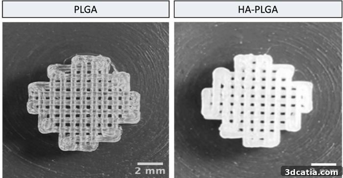
3D-printed bone structures (photo credit: Fatih Eroglu)
Specifically, the Henry Royce team prints porous bone-like scaffolds that serve as domiciles for cancer cells—primarily breast cancer cells—to observe how they colonize and interact with bone tissue. The researchers use two main materials to produce these scaffolds: PLGA and a composite referred to as HA-PLGA. PLGA (poly(lactic-co-glycolic acid)) is a biodegradable polymer commonly used in tissue engineering to form the structural backbone of a scaffold. HA-PLGA combines PLGA with hydroxyapatite, the mineral naturally found in bone, to create a matrix that more closely mimics the chemical and mechanical cues of real bone. By tuning the scaffold composition and architecture, the team can recreate a more realistic bone microenvironment for controlled laboratory studies of metastasis and cell behavior.
For fabrication, the group favors fused deposition methods—an accessible form of 3D printing that deposits melted material layer by layer. This choice substantially lowers equipment costs: the researchers report using a setup that cost under €1,000, rather than the significantly more expensive bioprinters that can cost many thousands of euros. The printed scaffolds act as three-dimensional frameworks that support cell attachment, proliferation and differentiation. Into these structures the scientists seed bone marrow-derived mesenchymal stem cells, a versatile cell type capable of differentiating into bone-forming cells. In these engineered environments, the stem cells not only survive but begin to create bone-like tissue, producing an in vitro model suitable for studying tumor–bone interactions.
Early experimental results have been encouraging. Cancer and stem cells adhere to the scaffolds, proliferate and, over time, show signs of differentiation toward bone-forming lineages. This outcome gives researchers a practical, humane platform to observe how metastatic cancer cells colonize bone tissue, respond to microenvironmental signals, and react to potential therapies. Such 3D-printed bone models can therefore serve as a valuable complement—or in some cases an alternative—to traditional animal testing, offering greater reproducibility, lower cost, and a reduced ethical burden.
“We are not just building scaffolds; we are creating new ways to study disease and test treatments that could reduce animal experiments while speeding research progress,” explains Fatih Eroglu.
The use of 3D-printed bone tissue models aligns with broader efforts in biomedical research to develop more physiologically relevant, ethically responsible, and cost-effective testing platforms. While such models do not yet replace all aspects of in vivo studies, they provide a powerful tool for screening hypotheses, studying cell–matrix interactions, and reducing the number of animals required for confirmatory experiments. Continued refinement of scaffold materials, printing resolution and biological protocols will likely enhance the realism and utility of these models for cancer biology, drug testing and regenerative medicine.
Photo credit: Fatih Eroglu / Henry Royce Institute
The post 3D-Printed Tumor Models Reveal How Cancer Cells Spread first appeared on 3dcatia.