Volumetric 3D Printing and Living Cells: Compatibility Timeline
This is the third major iteration by the team of scientists at the École Polytechnique Fédérale de Lausanne (EPFL) working to refine a volumetric 3D printing technique first developed in 2017. According to their latest publication, this updated method is roughly 70 times more efficient than earlier versions and, for the first time, enables the printing of structures that incorporate living cells.
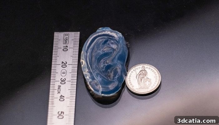
The most striking outcome of this new study is a life-size human ear printed from a gelatin-based hydrogel. The ear was printed with embedded human cells that, six days after printing, remained viable and had begun to organize into network-like structures—an important early indicator for tissue engineering and bioprinting applications.
Human ear printed using the TVAM process (photo credits: LAPD / EPFL).
What is TVAM?
The technique is called tomographic volumetric additive manufacturing (TVAM). In broad terms, TVAM builds three-dimensional objects by projecting light patterns into a rotating container filled with a photosensitive resin. By delivering light from multiple angles and carefully controlling the exposure, a full 3D object forms inside the resin volume in a matter of seconds to minutes—without the layer-by-layer approach common to traditional stereolithography or fused deposition modeling.
EPFL has been developing TVAM since 2017, when an initial team established the method’s basic principles. A later research group led by Professor Christophe Moser at the Laboratory of Applied Photonics Devices (LAPD) continued that work and published a series of three progressive improvements that systematically addressed the technique’s limitations for biomedical materials.
2022: At that stage, standard TVAM worked well only with transparent resins that allowed light to pass through without scattering. Many biomaterials and cell-laden resins are optically opaque or scattering, causing the projected light to deviate before reaching its target regions. To address this, researchers integrated a camera to monitor the actual light paths and applied real-time computational corrections. This approach reclaimed much of the precision previously achievable only with clear resins.
2025: Even with optical corrections, only a small fraction of the projected light—about 1%—actually reached the target volume in scattering biomaterials, forcing the use of very high optical power. The team then adopted a holographic projection strategy, projecting an engineered hologram directly into the rotating resin container. This improved light delivery efficiency dramatically, reducing required optical power by roughly 25-fold while improving printing outcomes for denser, scattering resins.
2026: The most recent advance replaces amplitude modulation hardware with a device that controls the phase of the light beam directly. Phase modulation allows precise shaping and steering of light through scattering media with far greater energy efficiency. This change increased overall process efficiency by approximately 70 times. With such a dramatic reduction in required optical energy, the team was able to use a modest 150 mW diode laser—substantially weaker than lasers used previously—yet still confirm cell viability after printing.
María Álvarez-Castaño, doctoral researcher, and Christophe Moser, director of the LAPD (photo credits: Adrien Buttier/EPFL).
Why the energy source matters
The switch in how light is modulated may sound like a technical detail, but it is central to making volumetric bioprinting compatible with living cells. Cells are sensitive to light exposure: too much light or the wrong wavelengths during printing can cause irreversible damage. By reducing the total optical energy delivered to the resin by roughly 70-fold, the research team succeeded in creating conditions under which true living cells survive the printing process.
This milestone—printing a tissue-scale structure that contains viable human cells—represents an important proof of concept rather than a finished medical product. The group at EPFL has outlined next steps that include improving projection fidelity, testing bioresins with higher initial cell densities, and refining the mechanical and biological properties of printed constructs so they more closely match target tissues.
Beyond this single demonstration, volumetric bioprinting with phase-controlled holographic projection opens new possibilities for rapid fabrication of complex, tissue-like structures. Faster print times, improved cell viability, and the ability to work with more optically challenging materials could accelerate research in tissue engineering, regenerative medicine, and organ-on-chip platforms.
The EPFL team’s work is a clear step forward in volumetric 3D printing and bioprinting. As projection fidelity and material formulations improve, the method may become a powerful tool for producing soft tissue constructs with embedded, living cells. Readers interested in advances in medical 3D printing, bioprinting, and volumetric additive manufacturing are encouraged to follow developments from academic laboratories and peer-reviewed publications to track progress and applications.
Cover photo: holographic projection of a human ear model onto a container holding a sample (photo credits: Adrien Buttier/EPFL).
The post Volumetric 3D Printing and Living Cells: Compatibility Timeline first appeared on 3dcatia.