ETH Zurich Develops 3D Printed Cardiac Patch
An interdisciplinary research team led by Professor Robert Katzschmann (ETH Zurich) and Professor Omer Dzemali (Zurich University Hospital) has unveiled a promising medical technology called the RCPatch. This 3D‑printed cardiac patch is designed to integrate with heart tissue, seal defects in the cardiac chamber and actively support tissue regeneration rather than remaining an inert foreign body.
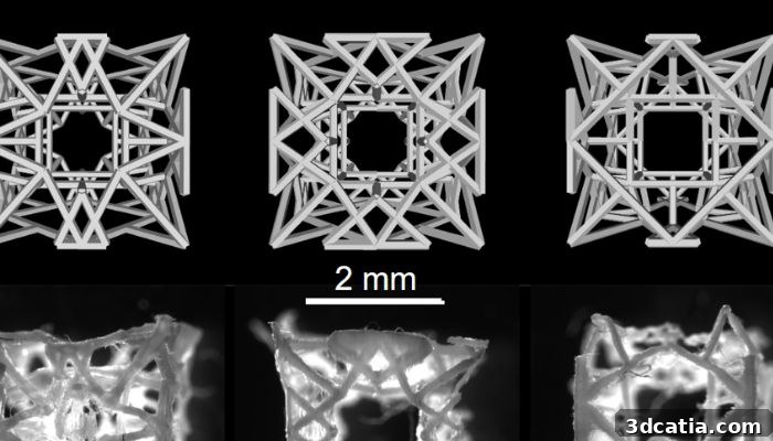
The lattice structure of the patch shown from three angles
A patch made of three coordinated components
The RCPatch combines three complementary elements: a fine‑mesh sealing net that directly covers the lesion, a 3D‑printed support frame formed from a biodegradable polymer lattice, and a tissue‑promoting hydrogel populated with living cardiac cells. The lattice structure is produced by 3D printing and filled with hydrogel, which is also applied to the sealing net. Once implanted, the patch is intended to integrate with adjacent heart tissue; over time the polymer support dissolves, leaving regenerated tissue in place rather than long‑term foreign material.
This integrated design aims to address several limitations of the current standard—bovine pericardial patches—which are mechanically stable and easy to implant but biologically inactive and permanent. Such permanent implants can be associated with complications like calcification, thrombus formation, or inflammation. By contrast, a biologically active, resorbable patch that supports native tissue growth could reduce those risks and improve functional recovery.
In preclinical tests using porcine models, the RCPatch successfully sealed an artificially induced defect in the left ventricle. Implantation proved straightforward, the patch withstood high blood pressures, prevented bleeding at the defect site and contributed to the restoration of cardiac function. These outcomes suggest clear advantages over currently used bovine pericardial patches, particularly when long‑term biological integration is the goal.
Promising outlook and next steps
The encouraging preclinical results provide a strong foundation for further development toward clinical use in humans. The research team envisions a mechanically reinforced, tissue‑based implantable cardiac patch for treating myocardial lesions. Achieving a clinically available product will, however, require extensive additional evaluation. According to the investigators, the immediate next steps include long‑term animal studies to assess overall device stability, durability under physiological conditions and the extent of true tissue regeneration over time.
Schematic illustrating how the patch is applied
If additional studies confirm safety and regenerative benefit, the RCPatch concept could mark a major step toward regenerative therapies for heart disease. Its combined focus on sealing defects, providing initial mechanical stability and enabling biological integration distinguishes it from passive repair materials. Longitudinal research will be required to verify sustained functional improvement, immunological compatibility and the timeline of scaffold resorption and tissue replacement.
Beyond the technical and biological challenges, translation to clinical practice will involve regulatory review, scaled manufacturing and standardized surgical procedures. Collaboration between bioengineers, cardiac surgeons and regulatory experts will be essential to transform the prototype into a device that can be evaluated in human clinical trials. For up‑to‑date details, consult the ETH Zurich research news and institutional publications describing the project.
The RCPatch illustrates how advances in 3D printing, biomaterials and cell‑based hydrogels are converging to create next‑generation cardiac repair options. Continued preclinical validation and careful clinical translation could make this approach a viable regenerative alternative to permanent, inert cardiac patches.
*Photo credits: Soft Robotics Lab / ETH Zurich
The post ETH Zurich Develops 3D Printed Cardiac Patch first appeared on 3dcatia.