How 3D Ice Printing Could Make Artificial Blood Vessels
The need for organ donations—particularly hearts, kidneys and livers—has become increasingly urgent, and many scientists are dedicating their work to finding ways to create viable artificial organs. Although progress has been uneven in recent years, a new approach from researchers at Carnegie Mellon University shows promise. By using 3D printing of ice, the team has developed a technique to fabricate structures that closely resemble blood vessels, mimicking natural vessel architecture and enabling functional behavior that could integrate with living tissue.
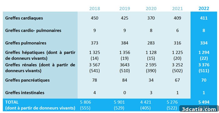
This development comes at a critical time. As of January 1, 2023, the Barometer on awareness and perception of organ donation in France reported that 10,810 patients were on active waiting lists—meaning they were immediately eligible for an organ transplant across all organ types. That number underlines the persistent gap between organ supply and demand and helps explain why advanced manufacturing methods such as 3D printing have attracted so much attention and hope within the biomedical community.
In 2022, transplants increased by 4% in France, yet many patients remain on waiting lists (photo credit: Agence de la biomédecine)
Many teams have explored techniques such as biofabrication and bioprinting to grow organs in the lab, but creating fully functional organs has been hindered by a major hurdle: building integrated networks of blood vessels that supply oxygen and nutrients throughout a tissue. Conventional artificial blood vessels and simple channel networks often fail to replicate the complexity, scale and surface properties of natural vasculature, limiting their ability to support living tissue over time. The 3D ice printing approach offers a different path to address that bottleneck by producing smooth, biomimetic channels that can be embedded in scaffold materials used for tissue engineering.
3D ice printing to create artificial blood vessels
The 3D ice printing method was originally developed by engineering researchers at Carnegie Mellon in 2022. The process involves depositing a controlled stream of water onto a very cold surface so that the water freezes layer by layer. In this specific implementation the researchers use heavy water—water in which hydrogen atoms are replaced by the isotope deuterium—because its higher freezing point and physical properties can produce a smoother frozen surface. Rather than freezing each deposited layer solidly, their technique keeps a thin liquid layer on top of the printed ice during fabrication. They call this a “freeform” process: by maintaining that liquid layer, the printed structures avoid the typical stepped layering texture of many additive methods and instead achieve a continuous, smooth geometry that is better suited for biomimetic channels.
Photo credit: Feimo Yang
After the ice templates are printed, they are embedded within a gelatin-based hydrogel known as GelMA, a material commonly used in tissue engineering because it can be crosslinked to form a stable matrix. The GelMA is then exposed to UV light to solidify the hydrogel while the ice templates melt away, leaving behind empty channels that mirror the printed vessel geometries. These channels can be seeded with endothelial cells—the specialized cells that line natural blood vessels—to create living, vessel-like conduits within the gel scaffold.
In their experiments, the Carnegie Mellon team successfully introduced endothelial cells into the printed channels and observed that the cells adhered and survived on the GelMA scaffold for at least two weeks. That initial survival demonstrates the potential of the technique to produce perfusable, cell-lined vessels suitable for longer-term culture and maturation. The researchers plan to continue cultivating these constructs for extended periods to study stability, cell organization and functionality over time.
Beyond the long-term objective of engineering transplantable tissues or organs, these realistic vascular networks could be highly valuable for preclinical testing. Engineered vessels embedded in hydrogels provide a platform for drug screening, toxicology assays, and studies of vascular disease under controlled laboratory conditions. Because the channels produced by the ice-printing approach can recreate complex branching patterns and smooth lumen surfaces, they may better replicate physiological blood flow and cell–matrix interactions than many traditional models.
While significant challenges remain—such as scaling up to organ-sized constructs, integrating multiple tissue types, and ensuring long-term perfusion and immune compatibility—the ice-based 3D printing method represents an innovative tool in the toolbox of organ engineering and vascular biofabrication. Its ability to generate smooth, biomimetic channels that support endothelialization is a meaningful step forward for efforts to bridge the gap between lab-grown tissues and clinically relevant grafts.
*Cover photo credit: Carnegie Mellon University
The post How 3D Ice Printing Could Make Artificial Blood Vessels first appeared on 3dcatia.