Simpath 3D Prints Realistic Tracheal Models for Medical Training
As we know, 3D printing has profoundly transformed the medical field, enabling the production of bespoke models ranging from anatomical replicas to fully patient-specific implants. One young startup working in this space is Simpath. Founded in 2023, Simpath develops realistic 3D models for anesthesia and respiratory medicine. Using modern 3D printing technologies, the company produces highly detailed anatomical structures that promise to improve medical simulation, training, and the development of new devices. In a conversation with the Simpath team, we learned how these models could shape the future of medicine and how their additive manufacturing process produces tracheal models.
3DN: Could you introduce yourselves and tell us how you started with 3D printing? How did Simpath come about?
The Simpath team combines clinical and design expertise: anesthetists James Broadbent and Jeremy Young, together with industrial designers Bernard Guy and Nicole Hone. Before founding Simpath, each team member had extensive experience with 3D printing in their own domain, gained through academic and research activities. Earlier projects—such as pediatric 3D-printed tracheas and work on pneumatic 4D printing techniques—laid the technical and conceptual foundations for Simpath. Our group formed through collaborative projects between universities and hospitals, where we discovered the potential of combining clinical insight with design and fabrication skills.
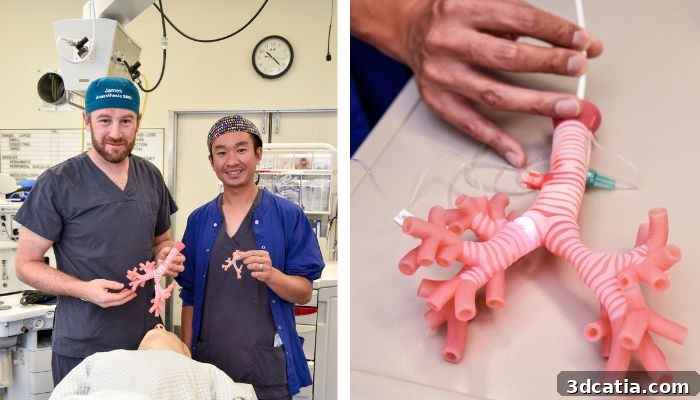
James Broadbent (left), Jeremy Young (right) and a close-up of the model.
Simpath was born from a clear goal: to improve training tools for airway management. We saw a significant gap in the market for anatomical models that are not only visually and tactilely realistic, but that also reflect the dynamic nature of the human body. Our aim is to build interactive models that bridge traditional physical models and emerging digital simulations, offering a more complete and practical learning experience.
A major strength is the close collaboration between clinicians and designers. Health professionals bring contextual expertise, identify clinical needs, and test prototypes, while our design team translates those needs into creative, practical solutions. This iterative partnership ensures that our models are both clinically relevant and well engineered for simulation and training.
3DN: What projects are you currently working on? What are the most notable anatomical models you’ve 3D printed so far?
We are currently developing a pediatric model designed for CICO training (Can’t Intubate, Can’t Oxygenate). This simulator includes a fixed head and neck combined with a flexible, interchangeable trachea so trainees can repeatedly practice emergency procedures under realistic conditions.
At the same time, we are enhancing a dynamic airway model that incorporates surrounding anatomical structures to increase realism. This adult trachea model reaches a level of fidelity not commonly found in physical trainers: it supports dynamic pathologies and innovative features that change the way airway management is taught.
Unlike static models or single-material parts, our trachea reproduces complex internal anomalies and dynamic behaviors such as distension, constriction, active bleeding, and arterial pulsation. This approach delivers an immersive, interactive simulation with meaningful visual and tactile feedback. That tangible realism gives physical models a decisive advantage over purely virtual simulations in many training scenarios.
Dynamic airway model with multiple realistic components.
Our models let clinicians rehearse complex procedures and emergency scenarios. For example, injecting fluid through a tiny orifice in the tracheal wall simulates active bleeding from a tumor. The design also supports precise surgical exercises—creating and draining a fluid-filled cyst, for instance. Users can simulate conditions like tracheal stenosis by inflating inner walls to create a narrowed passage, or reproduce bronchoconstriction and other respiratory pathologies for teaching and device testing.
3DN: Who uses your anatomical models and for what purposes?
Our models are primarily intended for surgical and medical training. Clinicians and students use them to practice procedures and prepare for real emergency situations. They offer an ethically acceptable way to practice difficult techniques and exploratory procedures with a level of realism that is hard to achieve otherwise.
They are also valuable for product testing and medical device development, where anatomically faithful conditions are essential for performance evaluation. Additionally, our models serve as educational tools for patient communication, helping patients and families better understand specific medical conditions and treatments.
The 3D model enables the simulation of a variety of emergency scenarios.
3DN: What materials and processes do you use?
Our design process emphasizes individuality, age, and health status. We move beyond generic static models toward data-driven simulations. To design anatomically accurate and device-compatible models, we use CT scans and 3D modeling tools such as Rhino and ZBrush. For fabrication, we rely on multi-material PolyJet 3D printing to achieve remarkably realistic textures. Using a mix of materials allows us to recreate a range of tissue consistencies from soft tissue to cartilage.
Clinical validation is an essential step in our workflow to ensure the models meet simulation needs. For example, our trachea model can visualize asthma or bronchoconstriction to explain why patients experience breathing difficulty and to demonstrate how bronchodilator treatments work.
Close-up of the tracheal model.
3DN: What challenges come with 3D printing anatomical models?
A major challenge is making models that are both realistic and functional. Unlike static models, our designs must reproduce dynamic conditions, which requires precise control over material composition and geometric structures to balance realism and performance. PolyJet materials can be fragile, especially in very small structures, so ensuring durability for repeated training use is another key concern. We continually refine materials and design to maintain realism without sacrificing robustness.
3DN: What are your long-term goals?
Building on our research published in Anaesthesia and Critical Care, we aim to develop comprehensive surgical training programs, high-fidelity simulation models, and physical anatomical representations that improve understanding for both clinicians and patients. High-resolution, full-color physical models provide rich information that complements emerging digital representations, which are still maturing and can be inconsistent.
Simpath’s 3D airway model.
We expect our models to play a central role in simulation centers, benefiting both learners and experienced professionals. There are also strong opportunities to collaborate with medical technology companies to test and evaluate devices. By staying at the forefront of 3D printing and incorporating continuous clinical feedback, we aim to drive innovation in medical education and ultimately improve patient care. To learn more or get in touch, visit Simpath’s website.
What do you think of Simpath’s work? Share your thoughts in the article comments. Interested in medical 3D printing news? Visit the original coverage here. You can also follow us on Facebook or LinkedIn.
*Photo credits: Simpath
The post Simpath 3D Prints Realistic Tracheal Models for Medical Training first appeared on 3dcatia.